Help4vet provides access to expert veterinary radiologists who offer interpretation and reporting services for X-rays of small animals (dogs and cats) on behalf of veterinarians.
We accept X-ray files in the DICOM (.dcm) format, and for each case, veterinarians can upload up to 15 images and select up to 3 areas of one patient. We offer two types of examinations: one that takes up to 48 hours and another that takes up to 72 hours. However, in most cases, our expert radiologists interpret the images within 3 to 10 hours, depending on the chosen option.
Our doctors meticulously examine the submitted images and prepare a detailed interpretation and report, which includes differential diagnosis based on the patient's history, clinical symptoms, and suggestions from the attending veterinarian. We also provide comments and guidance to attending veterinarians on how to improve photo quality if we observe any positioning or exposure parameter errors.
The completed radiography report is sent to the veterinarian's email address as a downloadable PDF file with comments in the system. At Help4vet, we aim to make veterinary radiology interpretation and reporting accessible, efficient, and accurate for our valued clients.
An X-ray machine consists of an X-ray tube head, a high voltage generator, and a control panel that includes a variety of regulators. The milliampere (mA) regulator adjusts the intensity of the X-rays produced, while the kilovolt (kV) regulator regulates the energy and penetration of the X-rays. In digital radiography (DR), X-ray transmission is electronically measured through the patient and then converted into a digital file for viewing on a computer workstation.
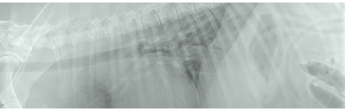
X-rays are primarily used to evaluate the skeletal system, such as fractures and degeneration, and soft tissues, including neoplastic changes, inflammations, swelling, and fluid in body cavities. They are especially useful for diagnosing post-traumatic changes, foreign bodies, urolithiasis, and gastric torsion, allowing veterinarians to make a preliminary diagnosis during a single visit. However, radiography has its limitations, as there is individual variation and great overlap between normal and mildly affected abnormal organs.
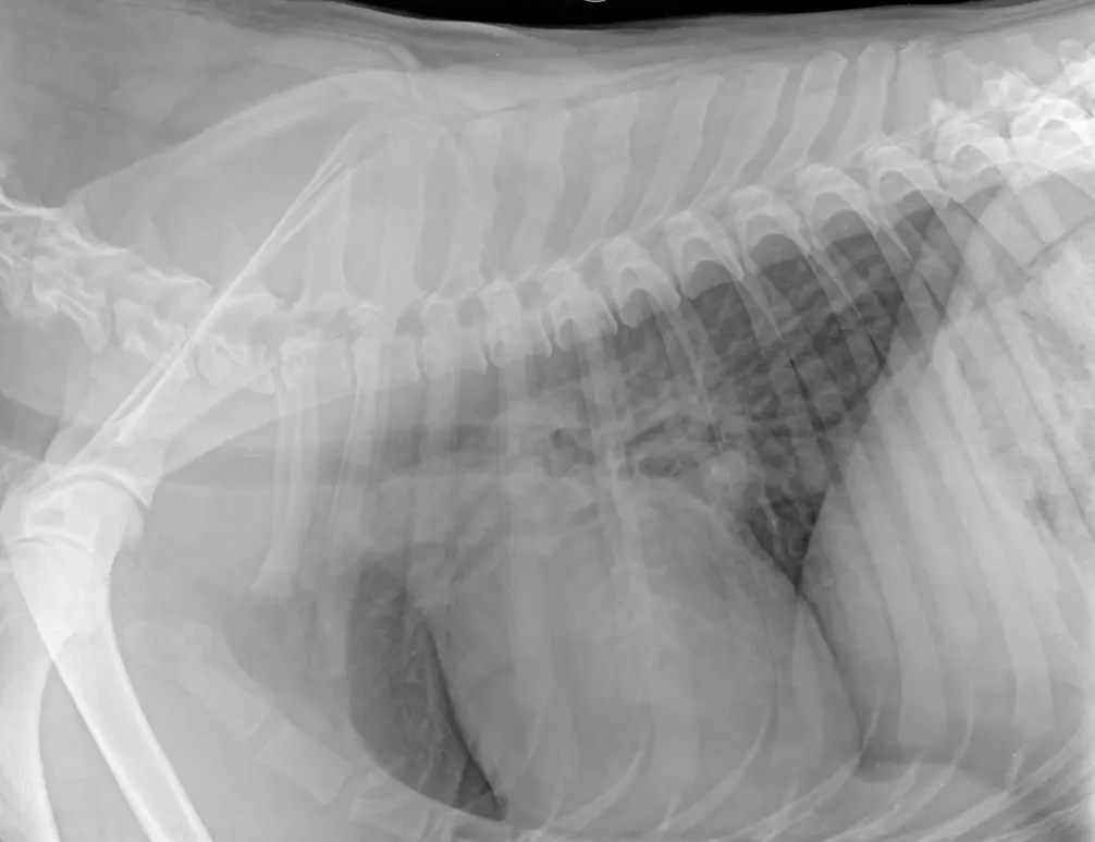
Although X-ray images are static, repeat or serial radiography performed over time can provide additional information. Radiography rarely provides information on function, but CT and magnetic resonance imaging (MRI) can demonstrate exquisite contrast in regions not well-served by radiography, such as the brain and spinal cord. The resolution of film–screen radiography is superior to that of CT and MRI, providing an overview of large regions of the body, such as the thorax and abdomen, as well as structural information on the skeletal system.
Our expert radiologists interpret X-rays to evaluate skeletal and soft tissue changes, providing a detailed report that includes recommendations for treatment. We also offer repeat or serial radiography for follow-up care, and we use CT and MRI when needed for more detailed imaging.
A radiologist who prepares reports has outlined the advantages of radiography over other radiology methods in veterinary medicine. Radiography allows for clear visualization of tissues with a wide range of attenuation, including bone and soft tissue, and thicker and thinner areas of the patient, all on the same image. The displayed images can be post-processed to increase diagnostic sensitivity, and digital images are easily stored, viewed, and transmitted. X-ray equipment is also more affordable than computed tomography, making it a profitable option for veterinarians.
The aim when taking a radiograph is to obtain an image that accurately represents the anatomy of the patient without distortion or unsharpness, which has good contrast and is of high quality and free of artifacts. Therefore, it is important to prepare the patient properly (clean coat of the animal, remove collars and harnesses, sometimes sedate the patient), correct positioning (radiolucent wedges, radiolucent troughs, sandbags, rope ties) and the correct exposure parameters. When selecting exposure factors, the 15% rule is helpful: By increasing the kV by 15%, the radiographic density of the film is doubled. In order to maintain the equivalent radiographic density, the mAs is halved (e.g. if you have an exposure of 50 kV and 10 mAs, increasing the kV to 57 and reducing the mAs to 5 results in similar film blackening). It is also significant to use markers to measure distance or mark sides.
Fluoroscopy is an increasingly used technique in veterinary clinics. It generates images using X-rays, but we see the result as an image in real time. An significant element is the C-arm, which can be rotated axially or sagittally around the table. This gives you the opportunity to evaluate the internal structure of the body and even the functioning of internal organs. Fluoroscopy may be necessary to show a recurrent.