Help4vet facilitates access to veterinary radiology experts, who interpret MRI of small animals (dogs and cats), for veterinarians.
We accept files only in DICOM (.dcm) format. For each case, you can upload up to 500 files, however we will provide report up to 4 areas of one patient per each case. We offer two types of examinations up to 48 hours and up to 72 hours. In practice, most of the cases are interpreted within 6 to 12 hours, depending on the option selected.
The ready interpretation of the radiograph is sent to your email address and also accessible in the system as a downloadable PDF file.
Magnetic resonance imaging (MRI) is an advanced imaging technique that is becoming increasingly popular in veterinary medicine. It enables, like computed tomography (CT), spatial visualization of the examined structures. Initially, it was mainly used as a scientific and research imaging modality, but with the increase in the number of specialists, as well as the increase in awareness and expectations of animal owners, it began to play a key role in clinical practice.
The big advantage of MRI, unlike CT, is the lack of use of ionizing radiation. MRI imaging is based on the phenomenon of nuclear magnetic resonance imaging, characteristic of the nuclei of some elements. The most commonly used element is the hydrogen atom, due to its significant occurrence in living organisms.
The general scheme of MRI function consists in placing the patient in a constant homogeneous magnetic field, with the examined part of the body placed within a special coil. The coil emits a radio frequency signal and receives a signal emitted back by the body. Then the emitted signal is processed by computer and the image is reconstructed.
Due to the magnitude of the magnetic field strength, MRI devices can be divided into high-field, medium-field and low-field strength. High-field devices allow for highly detailed images of tissues, but medium-field devices are considered to be the golden standard in clinical imaging.
Both MRI and CT allow for accurate imaging of such head structures as nasal cavities, frontal sinuses, or tympanic cavities and ear canals, but MRI as the only available imaging technique allows for full and detailed diagnosis of brain diseases, such as ventriculomegaly, inflammatory, developmental or neoplastic changes. MRI exposes the brain structures in three imaging planes and allows making accurate measurements of various structures, for example, to determine the size factor of the cerebral ventricles. Differences in the signal of individual tissues (depending on the degree of their hydration), as well as the possibility of intravenous administration of contrast agents (changing the signal of tissues in the event of a blood-brain barrier break) allow for accurate differentiation of the above-mentioned pathological changes.
MRI is considered the golden standard in the imaging of spinal disorders because it gives the possibility of spatial visualization of structures and diagnostics of changes in the meninges of the spinal cord and the spinal cord itself. A significant number of pathological changes occurring in the spinal area, such as intervertebral disc disease, can also be traced in CT, but conditions involving the spinal cord and surrounding meninges can easily be overlooked using this technique. MRI allows obtaining a myelography-like image thanks to the contrast between the cerebrospinal fluid signal and the subarachnoid space, without the need to provide a contrast agent.
MRI is also used in the diagnosis of some diseases of the musculoskeletal system. CT allows accurate imaging of the skeleton and joint surfaces, while MRI will be used primarily to detect diseases of the surrounding soft tissues. It makes possible to assess the structure of tissues based on the diversity of their signal, as well as to identify and classify injuries of muscles, tendons, and ligaments.
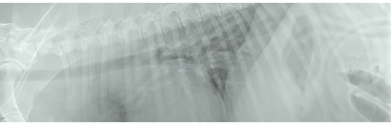
CT is used to assess the structures located in the abdominal cavity and chest, due to the poor contrast and sharpness of the image of these tissues, obtained in MRI.
To sum up, magnetic resonance imaging is an increasingly common imaging technique characterized by the ability to generate three-dimensional images of the examined structures and the lack of use of ionizing radiation, which is very important for the patients’ and staff’s safety. The ability to accurately image structures such as the brain, spinal cord or elements of the musculoskeletal system make MRI an excellent diagnostic tool, possible to use in everyday clinical practice.